Diagnostic Dermatology

Our Best Services

Diagnostic Dermatology

image

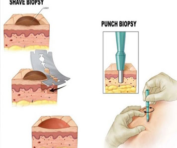
Skin biopsy- Shave/Excision

Skin biopsy is a procedure where a small sample of skin tissue is removed for examination under a microscope. This can help diagnose a variety of skin conditions, including skin cancers, infections, and autoimmune disorders.

image

Histopath reports

Histopathology reports are written by pathologists who examine the skin biopsy sample under a microscope. The report includes a description of the tissue, the type of cells present, and any abnormalities or changes that are seen. The report can help diagnose skin conditions and guide treatment decisions.

image

IHC markers

IHC markers (immunohistochemistry markers) are special stains used in pathology to help identify certain proteins or cells in a tissue sample. These markers can be useful in diagnosing skin cancers, identifying the type of cell involved in a skin condition, and determining the prognosis of certain skin diseases.

image

Dermoscopy/Trichoscopy

Dermoscopy is a non-invasive imaging technique that allows dermatologists to examine skin lesions using a special microscope. This technique helps dermatologists to diagnose skin cancers, melanoma, and other skin conditions.

image

KOH mount & Fungal culture

KOH mount and fungal culture are laboratory tests used to diagnose fungal skin infections. A KOH mount involves examining a skin scraping under a microscope after treatment with potassium hydroxide, while a fungal culture involves growing a sample of skin on a special medium to identify the specific type of fungus causing the infection.

image

Immunofluorescence

Immunofluorescence is a laboratory technique that uses fluorescent antibodies to detect specific proteins or antibodies in tissue samples. This technique can help diagnose autoimmune skin diseases such as lupus, pemphigus, and dermatitis herpetiformis.

Contact us today for Healthy, Glowing Skin!